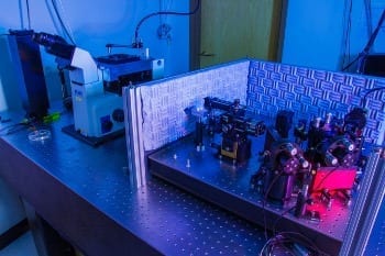
JILA instrument for accurately tracking microscopic objects such as DNA molecules for many hours. The microscope is on the left. The sample is mounted on the black block on top of the silver stage. The lasers and optics are on the right.
Credit: Burrows/JILA
JILA researchers have designed a microscope instrument so stable that it can accurately measure the 3D movement of individual molecules over many hours—hundreds of times longer than the current limit measured in seconds.*
The technology was designed to track the machinery of biological cells, down to the tiniest bits of DNA, a single “base pair” of nucleotides among the 3 billion of these chemical units in human genes. But the instrument could be useful well beyond biology, biochemistry and biophysics, perhaps in manufacturing.
JILA is a partnership of the National Institute of Standards and Technology (NIST) and the University of Colorado Boulder.
“This technology can actively stabilize two items relative to each other with a precision well below one nanometer at room temperature,” JILA/NIST physicist Tom Perkins says. “This level of 3D stability may start to interest the nanomanufacturing world, when they look at making and characterizing things on the single-nanometer scale.”
The work builds on JILA’s world-leading expertise in measuring positions of microscopic objects. The latest tweaks extend stability for a much longer time period, many hours at a time. With the longer observation times, researchers can see more successive steps of molecular motors, for instance. These biochemical processes are responsible for a broad range of movement in living organisms, including moving molecules around the interior of a cell or copying DNA into another form of genetic material, RNA. The new JILA instrument also can aid in measuring individual proteins as they fold into specific positions, a process required for them to work properly.
Until now, researchers had difficulty detecting more than a few individual, one-base-pair steps in succession before instrumental “drift” would blur the signal. Observing such sets of repetitive steps is very rare. The instrument must be stable to within about one-tenth of a nanometer (1 angstrom to biologists, equivalent to the diameter of a hydrogen atom).
Typically, a microscope can only occasionally achieve this level of stability. But when augmented by the new JILA measurement platform, it can reliably achieve tenth of a nanometer stability for up to 100 seconds at a time. And it can do this over and over again for extended periods—the JILA team operated the system for up to 28 hours straight.
In addition to its high precision and stability, the instrument can detect motion over a wide range of time scales, critical for calibrating instruments and measuring short-lived states in protein folding. The JILA method can be applied to optical trapping techniques, atomic force microscopes and super-resolution imaging.
Read more: Ultra-stable JILA Microscopy Technique Tracks Tiny Objects for Hours
The Latest on: Ultra-stable Microscopy
[google_news title=”” keyword=”Ultra-stable Microscopy” num_posts=”10″ blurb_length=”0″ show_thumb=”left”]
via Google News
The Latest on: Mltra-stable Microscopy
- Scientists pioneer new X-ray microscopy method for data analysis 'on the fly'on April 24, 2024 at 9:54 am
A new streaming technique allows playback of data while it is being generated. When scientists want to look at a tiny structure in a material, even one just a few atoms in size, they frequently turn ...
- electron microscopeon April 23, 2024 at 5:00 pm
When all you’ve got is a hammer, everything looks like a nail. And when you’ve got a scanning electron microscope, everything must look like a sample that would be really, really interesting ...
- Eugene Wigner’s Theoretical Electron Crystal Observed by Scientists for the First Timeon April 10, 2024 at 11:33 pm
Electrons, the enigmatic subatomic particles, are not solely confined to the orbits around atomic nuclei; they can also exist independently within the universe. Eugene Wigner, an eminent theoretical ...
- Super-Resolution Microscopy Can Be Super-Accessibleon April 9, 2024 at 9:26 am
To bring single-molecule localization microscopy to everyday researchers, laboratories can upgrade existing systems or deploy all-new platforms.
- Research team exerts electrical control over polaritons, hybridized light-matter particles, at room temperatureon April 9, 2024 at 6:41 am
integrating super-resolution microscopy previously invented by Prof. Kyoung-Duck Park 's team with ultra-precise electrical control. The resulting instrument not only facilitates stable generation ...
- An ultracompact multimode meta-microscopeon March 28, 2024 at 5:00 pm
Notably, the proposed guided-wave illumination module not only provides a low noise imaging mode, but also further reduces the system size that favors the compact microscope very much. As a result ...
- Center for Advanced Microscopy and Imagingon February 28, 2024 at 10:27 am
The Center for Advanced Microscopy and Imaging (CAMI) is an all-university research, teaching, and service facility located in Upham Hall on Miami University's main campus, in Oxford, Ohio. CAMI ...
- Zeiss Ultra Plus Field Emission SEMon November 15, 2023 at 3:50 am
The Ultra Plus scanning electron microscope is suitable for high-resolution imaging of biological and non-biological specimens. The microscope's charge compensation system allows non-conducting ...
- INFINITY3S-1UR – An ultra-sensitive, low noise microscope cameraon May 17, 2023 at 7:05 am
When versatility, ease of use, and cost are important decision factors, the Teledyne Lumenera INFINITY3S-1UR microscope camera is the best option. The INFINITY3S-1UR is an ultra-sensitive ...
- X-ray Microscopyon September 5, 2022 at 4:20 am
Rösner, Benedikt Finizio, Simone Koch, Frieder Döring, Florian Guzenko, Vitaliy A. Langer, Manuel Kirk, Eugenie Watts, Benjamin Meyer, Markus Loroña Ornelas ...
via Bing News


