via Wyss Institute
The work represents a major step toward a longstanding goal of tissue engineers: creating human tissue constructs realistic enough to test drug safety and effectiveness.
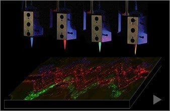
A new bioprinting method developed at the Wyss Institute for Biologically Inspired Engineering at Harvard University and the Harvard School of Engineering and Applied Sciences (SEAS) creates intricately patterned 3D tissue constructs with multiple types of cells and tiny blood vessels. The work represents a major step toward a longstanding goal of tissue engineers: creating human tissue constructs realistic enough to test drug safety and effectiveness.
The method also represents an early but important step toward building fully functional replacements for injured or diseased tissue that can be designed from CAT scan data using computer-aided design (CAD), printed in 3D at the push of a button, and used by surgeons to repair or replace damaged tissue.
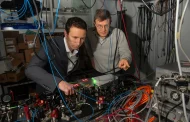
“This is the foundational step toward creating 3D living tissue,” said Jennifer Lewis, Ph.D., senior author of the study, who is a Core Faculty Member of the Wyss Institute for Biologically Inspired Engineering at Harvard University, and the Hansjörg Wyss Professor of Biologically Inspired Engineering at Harvard SEAS. Along with lead author David Kolesky, a graduate student in SEAS and the Wyss Institute, her team reported the results February 18 in the journal Advanced Materials.
Tissue engineers have tried for years to produce lab-grown vascularized human tissues robust enough to serve as replacements for damaged human tissue. Others have printed human tissue before, but they have been limited to thin slices of tissue about a third as thick as a dime. When scientists try to print thicker layers of tissue, cells on the interior starve for oxygen and nutrients, and have no good way of removing carbon dioxide and other waste. So they suffocate and die.
Nature gets around this problem by permeating tissue with a network of tiny, thin-walled blood vessels that nourish the tissue and remove waste, so Kolesky and Lewis set out to mimic this key function.
3D printing excels at creating intricately detailed 3D structures, typically from inert materials like plastic or metal. In the past, Lewis and her team have pioneered a broad range of novel inks that solidify into materials with useful electrical and mechanical properties. These inks enable 3D printing to go beyond form to embed functionality.
To print 3D tissue constructs with a predefined pattern, the researchers needed functional inks with useful biological properties, so they developed several “bio-inks” — tissue-friendly inks containing key ingredients of living tissues. One ink contained extracellular matrix, the biological material that knits cells into tissues. A second ink contained both extracellular matrix and living cells.
To create blood vessels, they developed a third ink with an unusual property: it melts as it is cools, rather than as it warms. This allowed the scientists to first print an interconnected network of filaments, then melt them by chilling the material and suction the liquid out to create a network of hollow tubes, or vessels.
The Harvard team then road-tested the method to assess its power and versatility. They printed 3D tissue constructs with a variety of architectures, culminating in an intricately patterned construct containing blood vessels and three different types of cells — a structure approaching the complexity of solid tissues.
Moreover, when they injected human endothelial cells into the vascular network, those cells regrew the blood-vessel lining. Keeping cells alive and growing in the tissue construct represents an important step toward printing human tissues. “Ideally, we want biology to do as much of the job of as possible,” Lewis said.
Lewis and her team are now focused on creating functional 3D tissues that are realistic enough to screen drugs for safety and effectiveness. “That’s where the immediate potential for impact is,” Lewis said.
Scientists could also use the printed tissue constructs to shed light on activities of living tissue that require complex architecture, such as wound healing, blood vessel growth, or tumor development.
The Latest on: Printing Living Tissues
[google_news title=”” keyword=”Printing Living Tissues” num_posts=”10″ blurb_length=”0″ show_thumb=”left”]
via Google News
The Latest on: Printing Living Tissues
- Coral reefs suffer fourth global bleaching event, NOAA sayson April 15, 2024 at 8:15 am
Along coastlines from Australia to Kenya to Mexico, many of the world's colorful coral reefs have turned a ghostly white in what scientists said on ...
- Will Heart Valve Tissue Engineering Change the World?on April 14, 2024 at 5:00 pm
Several strategies for heart valve tissue engineering are evolving ... to ensure a patient-specific immune tolerance to the living-tissue engineered valve. So far, the matrices developed have ...
- ERC Advanced grant awarded to crack the code of cartilage repairon April 12, 2024 at 10:52 pm
In our aging society, healing joint problems is becoming increasingly important. To do this, cartilage damage must become repairable.
- Can we crack the code of cartilage?on April 11, 2024 at 3:07 am
The best conditions for arches Jos links all the knowledge gained from research on other mammals and the models to his expertise in 3D bioprinting – printing living cells and tissues.
- The Hermiton April 10, 2024 at 5:02 pm
During the failed test, an "S" virus leak occurred. The virus has infected you, gradually killing the living tissues of your body. Hurry up to find an antidote and save the world from a global ...
- How organ, tissue and eye donations can save liveson April 5, 2024 at 2:46 am
April is National Donate Life Month, which creates awareness for lives that are healed and saved through organ, tissue and eye donation, and the need for people to participate in Utah’s organ ...
- Bioelectronic mesh capable of growing with cardiac tissues for comprehensive heart monitoringon March 21, 2024 at 5:00 pm
However, ways to effectively monitor living cardiac tissue are extremely limited. In part, this is because it is very risky to implant sensors in a living heart, but also because the heart is a ...
- A bioelectronic mesh capable of growing with cardiac tissues for comprehensive heart monitoringon March 20, 2024 at 5:00 pm
However, ways to effectively monitor living cardiac tissue are extremely limited. In part, this is because it is very risky to implant sensors in a living heart, but also because the heart is a ...
- Physiological Action of Acetic Acid in Living Tissueson March 12, 2024 at 5:00 pm
STARTING from the general thesis that volatile acids are produced in large quantity in the rumen of the sheep, McAnally and Phillipson 1 showed that some at all events of these acids are absorbed ...
- World's First 3D-Printed Neural Tissue Grows And Functions Like a Human Brainon February 5, 2024 at 7:46 pm
and other components to build living structures – has huge potential for creating tissues that replicate, and in some cases even replace the real deal. "Because we can print the tissue by design ...
via Bing News


